Treatments
Surgery is the first line of treatment for an ependymoma...
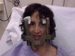
Picture above is IMRT radiation at Inova Fairfax Hospital
Overview
Surgery is the first approach in treating an ependymoma of the brain. The difficulty is not harming the patient in the process of removing the tumor. The typical location of an ependymoma (fourth ventricle with brain stem involvement) is the reason this tumor can be devastating to remove. It's tough to remove an entire tumor without damaging healthy brain tissue - this is where the skill of the neurosurgeon comes in. One surgeon explained it like this: "The risk is the surgeon doesn't know where the tumor ends and the brain stem begins til he gets there. It's like digging for an egg in the bottom of a sandbox with a big shovel and you don't know how deep the sand is, and you don't know where the egg is until you've broken it." Sometimes an ependymoma is so large that it is blocking the flow of cerebro-spinal fluid (CSF), a situation that in and of itself can be deadly. Debulking the tumor, and hopefully achieving a gross total resection if possible, relieves the intracranial pressure caused by the tumor. A shunt also can be placed to relieve the pressure (I had this done on 10/4/2005). The term "craniotomy" refers to the surgery and the removal (and replacement) of bone in the skull to reach the tumor. One benefit of surgery is that a biopsy can be performed, thus determining the exact grade and cell type of the tumor tissue - these results often guide further treatment.Radiation usually follows surgery to remove an ependymoma, though some patients do not have it after a first surgery. Radiation oncologists strive to find the "perfect" amount of radiation - to kill any remaining microscopic tumor cells but to avoid causing permanent damage to the healthy parts of the brain. With adults, this is fairly straight-forward; with children, it's another matter. It is not advisable for young children to receive radiation to their brain. Radiation is most often given over a period of time, or in "fractions" - this is the conventional radiotherapy that most ependymoma patients receive post-operatively to bathe the general area of the tumor bed with radiation. Stereotactic radiosurgery is a single, high dose of radiation targeted to a specific spot. When surgery is no longer possible, and conventional radiation has been used, adult ependymoma patients might undergo this kind of radiation. Logically, with this higher, more lethal dose, the possibility for side effects is greater. "Gamma knife" is the most well-recognized stereotactic radiosurgical technique. "Cyber knife" is another newer technique that delivers a high dose of radiation to the tumor but over several days rather than in a single treatment. One advantage of cyber knife is that the patient doesn't have to wear the head frame that is necessary for gamma knife. Radiation has serious risks and definite side effects that for me seemed to build up over time. There is the risk of damaging the vital brain tissue as well as the risk that the target tumor will swell afterwards. The effect is similar to a growing tumor and the swelling would have to be controlled with steroids.Chemotherapy promises to be a positive step in fighting glial tumors though there are few studies specifically carried out on ependymomas. Hopefully new techniques will be developed to allow the cancer-killing drugs to break through the so-called "blood-brain barrier" - the barrier that protects brain cells from allowing foreign particles to get through. (Interestingly, oncologist Paul Zeltzer wrote in his comprehensive book Leaving the Garden of Eden (2006) that the blood-brain barrier is not the major problem; rather, it's the lack of drugs available.) Temodar, made by Schering,(www.temodar.com) is a leap forward in treating higher grade glial tumors such as astrocytomas. Unfortunately, my ependymoma did not shrink or stop growing after two cycles of Temodar. My doctors tell me that drug therapy is improving rapidly and the longer I hang in there, the better my chances to find a cure. The National Brain Tumor Foundation has published a good overview of new chemotherapy categories - growth factor inhibitors, cell signal transduction modulators, anti-invasion agents, differentiating agents, anti-angiogenic drugs and cytoxic agents.
My Treatments
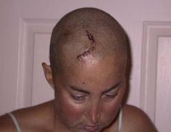
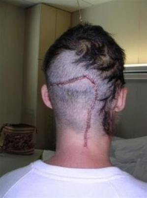
Surgery on May 3, 2000I was operated on by Laligam N. Sekhar, MD at George Washington University Hospital in Washington, DC. My tumor was nearly two inches long and was firmly attached to my brain stem. I was hospitalized for nearly 7 weeks and went to in-patient rehab for 10 days. I had several setbacks after surgery which prolonged my hospital stay. I had emergency abdominal surgery after doctors suspected that my PEG tube (a feeding tube inserted because I could not swallow) had been placed incorrectly. I had a tracheotomy and learned to speak with a Passe Muir valve. I also had post-operative pneumonia. I spent several weeks in the ICU and was looked after round-the-clock by my incredible family and friends who were my advocates when I was my sickest. I lost nearly 25 pounds in the hospital.Surgery on March 2, 2004My recurrent tumor was first seen on my annual MRI in April of 2003, then was followed closely until finally it was seen as a full-blown recurrence and Dr. Sekhar operated again. He predicted I would have terrible deficits, including a permanent tracheostomy, but my tumor was relatively easy to remove and there was no damage to my cranial nerves as feared, thanks both to Dr. Sekhar's skill and to the monitoring of my 10th cranial nerve in particular. Unfortunately, though, Dr. Sekhar felt the tumor was malignant and refers to it as an "aggressive ependymoma." He did not remove the entire tumor. He removed all he could see under the microscope, but residual tumor was apparent on the post-operative MRI. Sometimes it is hard to differentiate between tumor cells and healthy tissue in the lining of the ventricles. Almost miraculously (and perhaps because of the fact that he did not affect as much tissue compared to the experience I had in 2000), I was discharged from the hospital on my birthday, one week after surgery. The pathology report read: "The tumor is an ependymoma, WHO grade II. By report, Ki-67 immunohistochemistry demonstrated a labeling index of 5-10%, which could imply a greater likelihood of recurrence."Radiation 2004Dr. Sekhar recommended I go to Inova Fairfax Hospital for my radiation. I was lucky to be under the care of the chairman of radiation oncology, Dr. Glenn Tonnesen.Here is Dr. Tonnesen's summary of my treatment:"We felt that the entire posterior fossa was at some risk for recurrent disease, although the area of residual enhancement adjacent to the surgical site appeared to be the area of highest risk. Treatment was begun between 4/8 and 5/17/04, using a four-field conformal technique to irradiate the cerebellum, brain stem, and the surgical wound with a dose of 5040 rad in 28 fractions (treatments). Then between 5/18 and 5/24/04 we continued treatment of just the abnormally enhancing area, using very small arc portals to deliver an additional 900 rad in 5 fractions for a cumulative dose to the high risk area of 5940 rad."To begin radiation, I had to have my mask formed and then go through two simulations to ensure the radiation would be entering my head in the right places. The mask is bad, but it can be tolerated. A good friend who is a radiation oncologist told me to look at the mask as a "friend" because it allows for accurate, pinpoint treatment. I recommend taking something such as Valium before the mask fitting and the simulations because wearing that mask for an extended period was claustrophobic. I actually took 2.5 mg of Valium every day before treatment just to make things go a little easier - by the end I was used to the mask, but I didn't want to risk having a panic attack so I kept up with the medicine. With all the medicines I have had to take, a little Valium once a day for six weeks did not harm me and it enabled me to cope with the treatments!Chemotherapy 2005The first time I saw Dr. Howard Fine at the National Cancer Institute was June 30, 2004. He did not recommend chemotherapy then but advised that I be scanned every two months. He explained that chemotherapy would "target your body more than it would target your tumor." (I couldn't help but remember these words when I began the heavy-duty chemotherapy drugs in March 2005.) At the time of our first appointment, Dr. Fine was hopeful that the radiation I had would stunt the growth of the remaining tumor. Unfortunately, it did not. In fact, the tumor did not seem to mind the radiation at all.VP Shunt October 4, 2005What a bizarre procedure! A catheter was placed in my head and it winds its way down to my intestine. This cured my hydrocephalus. I was only in the hospital for two nights.Surgery on December 13, 2005I was unsure about another surgery. I was told several opinions about its safety and efficacy. All doctors said it would not be a cure but it would certainly be a way to remove some of the tumor but at an unknown cost. Dr. Jane and I were a good match and I felt I was in very good hands. I also liked the fact that by taking the risk, I was doing something rather than waiting to die.Just like I have done for all my surgeries, I waited with wonderful Margaret in the holding area until they wheeled me away - she is such a comfort. There's really no way to describe the sick feeling before you know you're about to undergo risky brain surgery. I smiled a lot and probably talked a lot to calm my nerves! I don't know how the surgical teams do it time after time, day after day, but I have always felt that the people in my OR and in the prepping area are heroes - they treated me like I was their only patient - their most important one ever. I don't know if these folks ever truly realize the impact they have because we never can go back and thank them - this is the nature of the OR experience.A combination of a miracle, Dr. Jane's expertise, and my abilities as a veteran patient (smile!) resulted in a terrific surgical result. It came at a cost however. But it was clear I wasn't going to survive without it and the damage that I could have suffered was horrible to consider. So coming out of it as intact as I did is an incredible outcome. Now if I can achieve an acceptable level of rehabilitation and also stay in remission for a long long time, that would be the best possible outcome. An excerpt from Dr. Jane's surgical report (12/13/05): We then brought the STEALTH system back into play and approached the tumor via lifting the tonsils and moving up the obex of the fourth ventricle. The tumor was readily encountered. Much to our surprise, there was a clear plane between tumor and floor of the fourth ventricle. We were able to go under the operating microscope and achieve a gross total resection... frozen specimen confirmed recurrent ependymoma.Gamma Knife Radiosurgery on February 18, 2013By Dr. Jason Sheehan to attack a recurrent tumor that was measurably growing in size over the course of six months - year.[2013]


